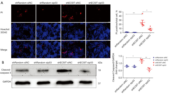
Figure 4.
ECSIT increases cell death dependent on p53 in MDA-MB-231 cells. (A) Cell death was detected by immunofluorescence of PI (red) and Hoechst 33342 (blue). Representative images (scale bar =50 µm) and quantification of PI-positive cells are shown (right panel; n=5). (B) Western blot analysis of cleaved caspase-3 and GAPDH in total lysates. Statistical significance was calculated for the indicated paired samples with *, P<0.05 and **, P<0.01. shRandom, negative control; shECSIT, knockdown of ECSIT; PI, Propidium iodide; siNC, negative control; sip53, using siRNA to suppress the expression of p53.

