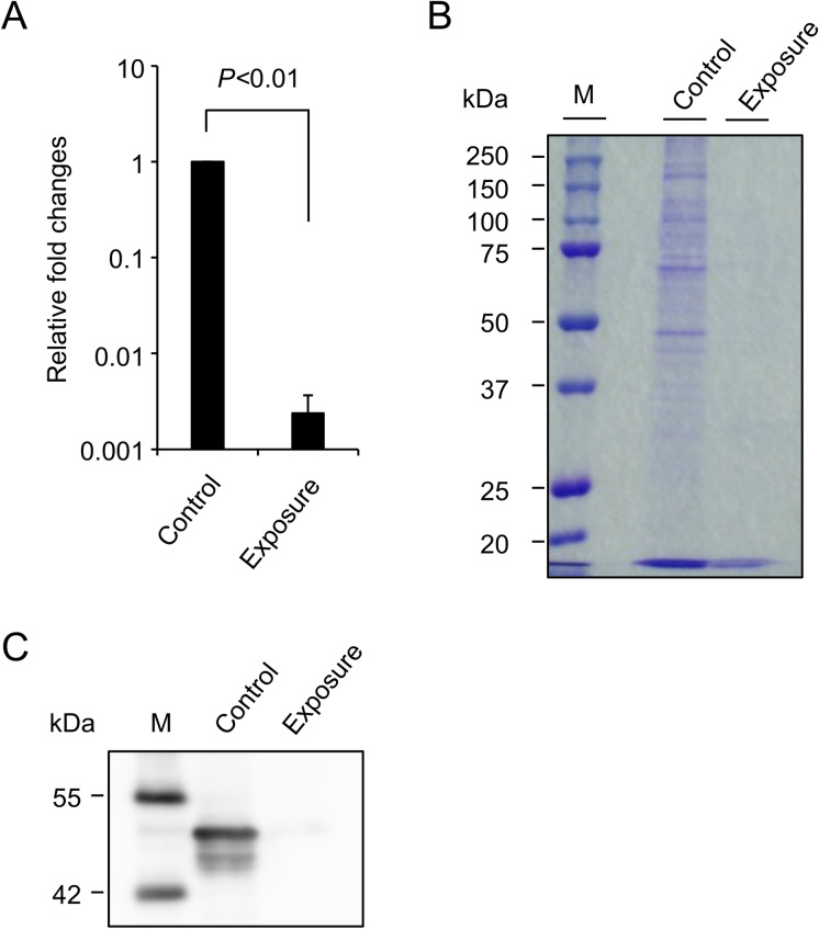
Fig. 5.
Damages to viral RNA and protein by NEAWPs. Concentrated virus was treated with (exposure) or without (control) NEAWPs for 1 h. (A) RT-qPCR was performed using 300 ng of RNA in each sample to amplify the SARS-CoV-2 nucleocapsid genomic RNA. The Y-axis indicates fold changes in the amount of nucleocapsid gene in NEAWPs-exposed group relative to that in the control group. All data represent the means + SD from three independent experiments. (B) (C) The same volume in each sample was loaded to gels and subjected to SDS-PAGE followed by CBB staining (B) or western blotting to detect the nucleocapsid protein (C). M indicates molecular markers. Images are representative of two (B) or three (C) independent experiments

