. 2018 Dec 10;8(72):41192–41206. doi: 10.1039/c8ra09506j

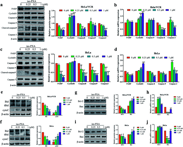
Fig. 6. Expression of apoptosis-related proteins and genes in HeLa/VCR and HeLa cells treated with iso-PXA. (a) and (c) After cells were treated with different concentrations of iso-PXA for 24 h, the expression of apoptosis-related proteins was determined by western blot, with β-actin as a loading control. Band intensities of western blots were quantified. (b) and (d) The mRNA level of cells treated with 0, 0.25, 0.5 and 1 μM iso-PXA for 24 h by RT-qPCR. Quantitative gene expressions were normalized to GAPDH. (e) and (f) After cells were treated with different concentrations of iso-PXA for 24 h, the expression of Bid and cytochrome c was determined by western blot, with β-actin as a loading control. Band intensities of western blots were quantified. (g) and (i) Western blot analysis of the expression of Bax and Bcl-2 proteins in HeLa/VCR and HeLa cells treated with iso-PXA (0, 0.25, 0.5 and 1 μM) for 24 h. Band intensities of western blots were quantified. (h) and (j) The levels of apoptosis-related genes in HeLa/VCR and HeLa cells treated for 24 h were analyzed by RT-qPCR. GAPDH was used as a loading control. Bars represent the mean ± SD (n = 3). *p < 0.05, **p < 0.01, ***p < 0.001 vs. the control group.

Fig. 6