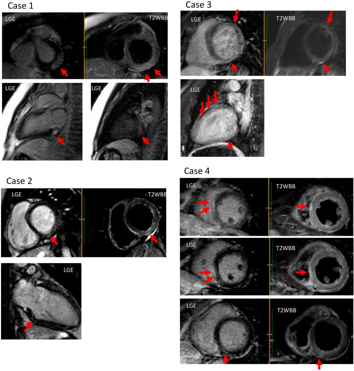
Figure 1.
Cardiac magnetic resonance imaging (MRI) of all profiled cases. Case 1: T2 high signal intensity and late-gadolinium enhancement (LGE) were observed at the sub-epicardial wall of the basal-mid inferolateral left ventricular (LV). Case 2: T2 high signal intensity and LGE were observed at the mid wall of the basal inferior LV. Case 3: T2 high signal intensity and LGE were observed at the mid wall of the anterior, and at the inferior LV. Case 4: T2 high signal intensity and LGE were observed at the mid-wall of basal inferior, and at the sub-epicardial wall of the mid- and inferoseptum LV.

