. 2018 Dec 6;8(71):40894–40911. doi: 10.1039/c8ra07163b

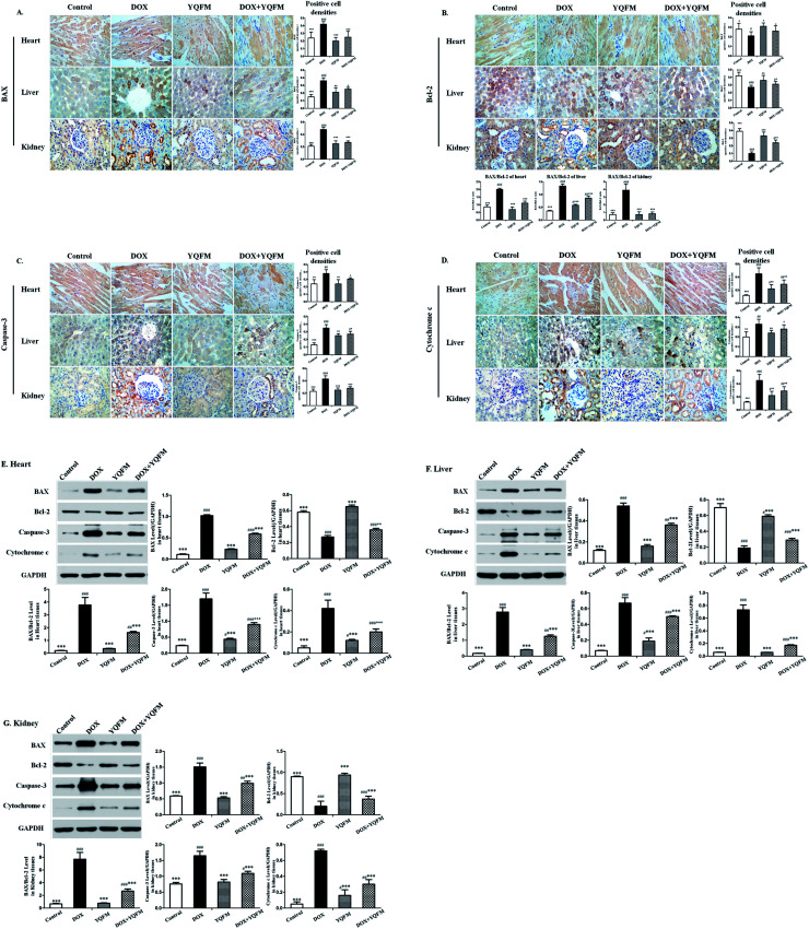
Fig. 11. YQFM remarkably ameliorated the expression of apoptosis-related proteins. Expression of apoptosis-related proteins (A) BAX, (B) Bcl-2, (C) caspase-3 and (D) cytochrome c by immunohistochemical staining (400×). Quantitative image analysis for immunohistochemical staining expressed as positive cell densities ratio across 10 different fields for each rat section in different tissue. Immunoblot and bar charts showed quantitative results for the expression of BAX, Bcl-2, caspase-3 and cytochrome c in (E) heart, (F) liver and (G) kidney. The results were shown as the mean ± SEM (n = 10) *p < 0.05, **p < 0.01, ***p < 0.001 vs. DOX group; #p < 0.05, ##p < 0.01, ###p < 0.001 vs. control group.

Fig. 11