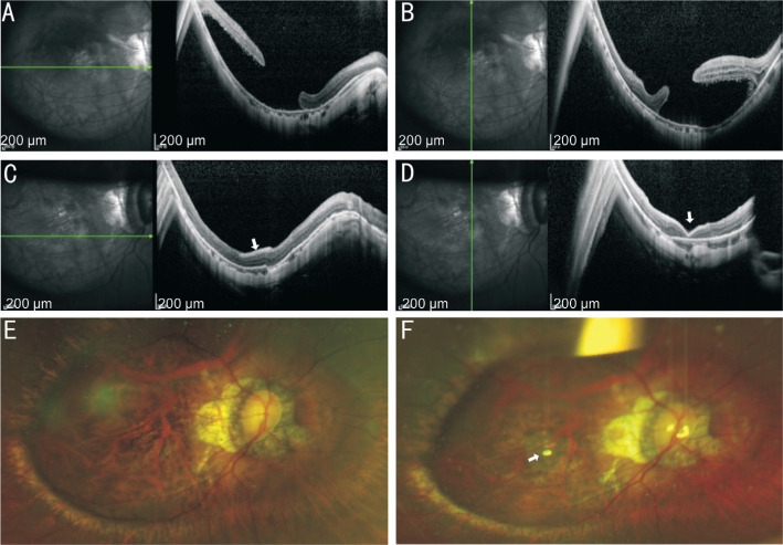
Figure 2. Information of the No.5 patient.
A, B: The OCT of macular area before operation showed the failure of MH recovery with macular regional RD; C, D: The OCT of macular area after operation showed that RD was reattached and MH was healed by covering AM (white arrow). E: The SLO before operation showed the failure of MH recovery with macular regional RD; F: The SLO after operation showed AM (white arrow) covered the MH. RD: Retinal detachment; MH: Macular hole; SLO: Scanning laser ophthalmosocopy; AM: Amniotic membrane.

