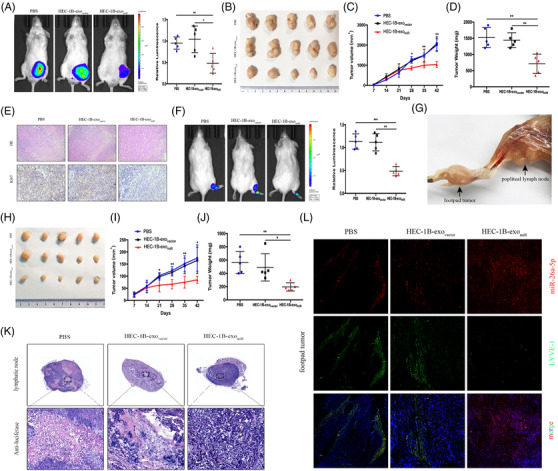
FIGURE 2.

Exosomal miR‐26a‐5p inhibits EC tumour proliferation and LNM in vivo. (A) Representative bioluminescence images and histogram analysis of subcutaneous tumours in NOD‐SCID mice, which treated with PBS, HEC‐1B‐exovector, or HEC‐1B‐exomiR (n = 5). (B) Images of subcutaneous tumours in mice treated with PBS, HEC‐1B‐exovector, or HEC‐1B‐exomiR (n = 5). (C, D) The tumour volumes and weights (n = 5). (E) Representative HE and immunohistochemical staining images demonstrating Ki67 expression. (F) Bioluminescence images and analysis of popliteal metastatic lymph node in NOD‐SCID mice, which treated with PBS, HEC‐1B‐exovector, or HEC‐1B‐exomiR after HEC‐1B cell inoculation into the footpad (n = 5). (G) Representative image of the popliteal lymph node. (H) Representative images of footpad tumours. (I, J) The measured footpad tumour volumes and weights (n = 5). (K) IHC of anti‐luciferase for popliteal lymph node treated with PBS or the indicated exosomes. Luciferase‐positive tumour cell in lymph node indicated metastasis. (L) Staining of miR‐26a‐5p and LYVE‐1 mice footpad tumour sections. Mean ± SD are provided. *p < .05, **p < .01, ***p < .001
