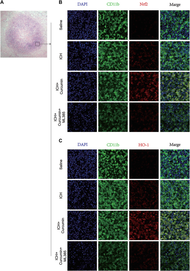
FIGURE 8.
Immunofluorescence staining of microglia in the surrounding hematoma at 7 days post ICH. (A) H&E staining in the tissue surrounding the hematoma. (B) The microglial membrane protein CD11b was labeled with FITC, and the membrane protein Nrf2 was labeled with Texas Red. (C) The microglial membrane protein CD11b was labeled with FITC, and the membrane protein HO-1 was labeled with Texas Red. n = 3/group. The data are from at least three such independent experiments.

