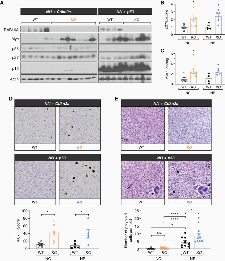
Figure 4.
Sustained RABL6A loss leads to paradoxical molecular and pathological alterations indicative of increased malignancy. (A) Representative western blots confirming loss of RABL6A, p16, and p53 in respective conditions. Rabl6 KO mice displayed increased p27 and Myc protein expression in both Nf1 + Cdkn2a (NC) and Nf1 + p53 (NP) tumors. ImageJ quantification of (B) p27 and (C) Myc protein expression. (D) Representative Ki67 IHC images (200X magnification) and H-score quantification reveal Rabl6 KO tumors have enhanced proliferation compared to WT tumors in both NC and NP settings. KO tumors notably also have enlarged cells. (E) Representative H&E images (scale bar, 75 µm) of the indicated tumors reveal increased cancer cell atypia with scattered enlarged cells in Nf1 + p53 altered tumors, which was further enhanced by RABL6A loss. Arrows highlight scattered enlarged cells with large, misshapen and multiple nuclei (morphologically consistent with polyploidy cells), which are magnified in the insets. Below, the number of enlarged cells with polyploidy was quantified and averaged from 5 different fields per tumor. B,C,D,E: Error bars, SEM. B,C,D: P value, Student’s t-test for comparisons between WT and KO tumors for the indicated tumor genotype (*, P < .05). E: P value, Tukey’s multiple comparisons test (*, P < .05; ****, P < .0001).

