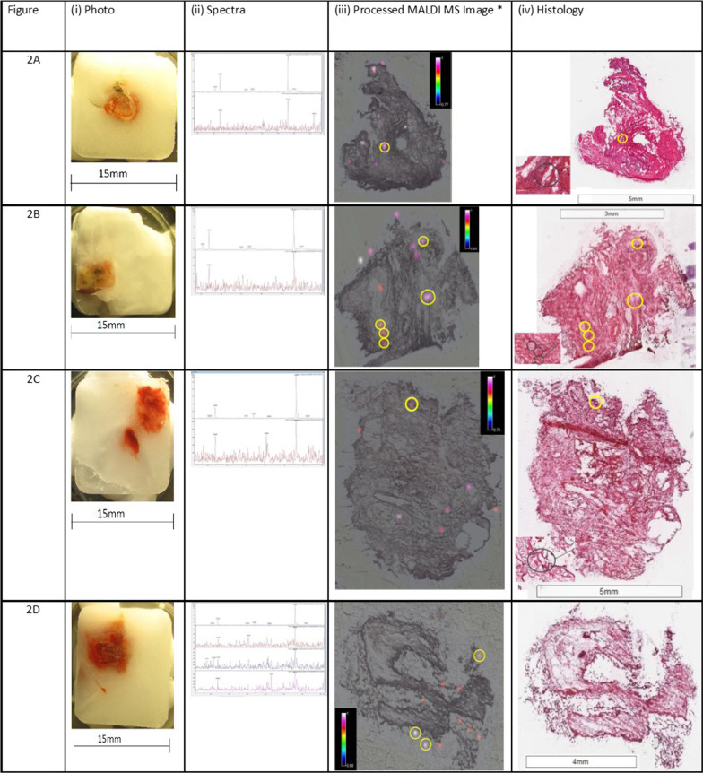
Fig. 3.
Representative MALDI-MS images, histology images and MS/MS for each participant. (TBC Samples). Each representative figure depicts the MALDI-MS image (100 µm pixel size) for the biopsy sample section and its corresponding histology image, a photograph of the frozen embedded biopsy sample and mass spectra showing both fragment ions (at m/z 123.9 and 166.0), obtained at the site of confirmed ipratropium detection (referred to as ipratropium or drug foci). For clarity, the MALDI-MS images for the detection of ipratropium have been adapted and the drug foci regions circled that are above the signal to noise threshold ratio 3:1 for both fragment ions (at m/z 123.9 and 166.0). The approximate location of these foci has been circled on the corresponding histology image

