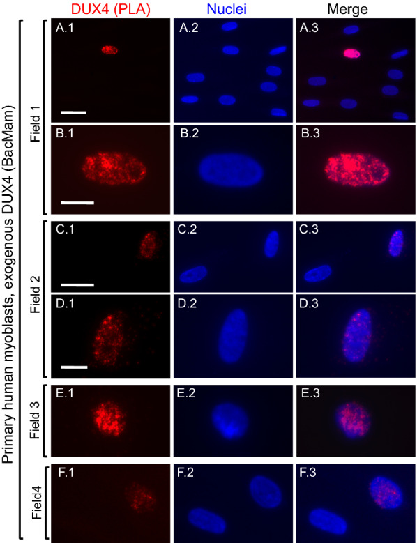
Fig. 1.

Validation of proximity ligation assay (PLA) for DUX4. Cultures of unaffected, primary human myoblasts were incubated with BacMam-DUX4 at a multiplicity of infection that generated DUX4 expression in a small proportion of the myoblasts. At 48 h after BacMam addition, cultures were processed for two primary antibody PLA (red signal) as described in Methods. Row A shows a single nucleus with a positive PLA signal (red) amidst eight nuclei that were unstained; and Row B shows the same nucleus at higher magnification to emphasize the characteristic punctate staining pattern expected for a PLA signal. Nuclei were stained with bisbenzimide (blue). Row C shows one nucleus with a positive PLA signal and a nearby unstained nucleus; and row D shows the positive nucleus at higher magnification. Rows E and F show additional examples of nuclei with positive PLA signals. Bar in A1 = 30 µm for row A. Bar in B 1 = 10 µm for row B. Bar in C1 = 25 µm for row C. Bar in D 1 = 10 µm for rows D, E, and F
