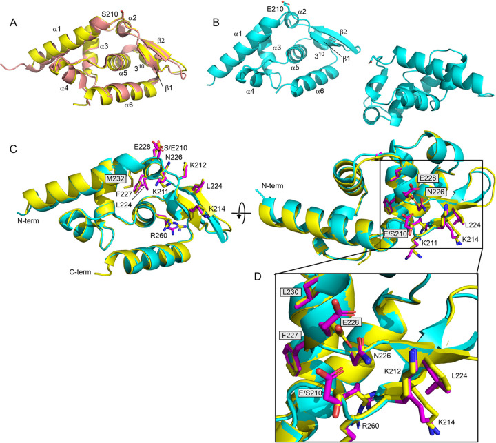
FIG 1.
Structural impact of phosphomimetic mutation S210E on Nishigahara PCTD. (A) Overlay of the wild-type PCTD from Nishigahara (yellow) and from CVS (salmon; 1VYI). Small structural differences are observed, particularly where Ni-PCTD has a shorter N terminus. Elements of regular secondary structure are labeled. (B) The two monomers of the crystal unit for S210E PCTD. (C) Overlay of the wild-type PCTD from Nishigahara (yellow) and S210E (cyan). The panel on the right is a rotation of 90° around the x axis. Side chains of the C-NES (L224, F227, L230, and M232) and the C-NLS (K211, K212, K214, and R260), as well as the site of mutation (S210E), are shown for wild-type (yellow) and S210E (magenta) PCTD. The change in interaction of N226 for E228 in wild-type (yellow) for E210 (magenta) in S210E is highlighted in the expansion in panel D.

