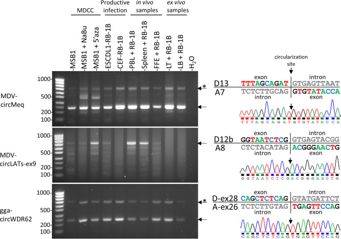
FIG 1.
Expression profiles of viral and cellular circRNAs in MDV infected cells that represent the different stages of viral infection. Agarose gel electrophoresis of inverse RT-PCR products obtained from two viral (meq and LATs) and one cellular (WDR62) gene are presented on the left part. Corresponding chromatogram results of the sequenced back-junctions obtained from the major amplicons are presented on the right. Both the exonic and intronic sequences of the donor and acceptor splice sites are indicated. MDCC: Marek’s disease chicken cell; FFE (Feather follicle epithelia); LT: ex vivo infected T lymphocytes; LB: ex vivo infected B lymphocytes; Asterisks (*) indicate rolling circle amplicons. Ladder: SmartLadder SF 100 bp-1kb (Eurogentec).

