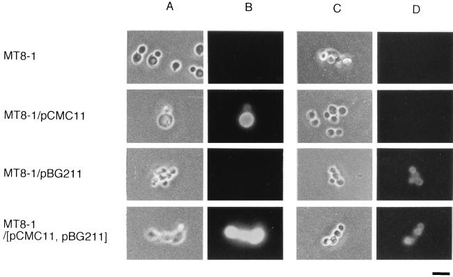
FIG. 2.
Immunofluorescence labeling of transformed cells. Phase-contrast micrographs (A and C) and immunofluorescence micrographs (B and D) of S. cerevisiae strains MT8-1 (host strain), MT8-1/pCMC11, MT8-1/pBG211, and MT8-1/pCMC11/pBG211 (MT8-1/[pCMC11, pBG211]). The primary antibody against A. aculeatus FI-CMCase (A and B) or the antibody against A. aculeatus β-glucosidase (C and D) was used. FITC-conjugated goat anti-rabbit IgG was used as the second antibody. Bar = 5 μm.

