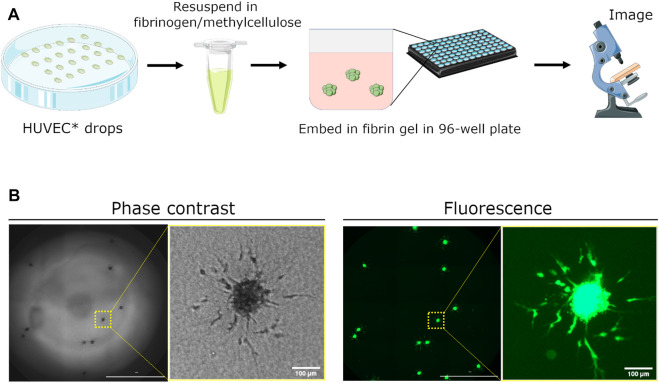
FIGURE 1.
Schematic demonstrating setup of spheroid assay for angiogenesis sprouting. (A) Human umbilical vein endothelial cells (HUVEC) are fluorescently dyed with CellTracker Green CMFDA (5 μm) and seeded as hanging drops for 24 h. Spheroids are collected, resuspended in a fibrinogen/methylcellulose solution, and mixed with thrombin in a 96-well plate to embed spheroids within the generated fibrin gel. Medium containing pro- or anti-angiogenic factors is added 1 h later to initiate sprouting. Angiogenic sprouting is imaged 24 h later using a real-time, fluorescence microscopy plate reader. (B) Representative images of embedded spheroids in a single well and a close-up of sprouting from a single spheroid. Images were acquired using phase contrast and fluorescence microscopy. Scale bar of whole-well image = 2000 μm.

