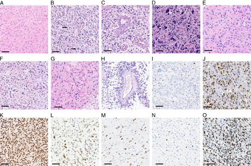
FIGURE 2.

Histomorphology and immunohistochemistry in H3 K27M-mt DMGs. A, Diffuse infiltrating glioma, conventional histologic grade 2. B, Diffuse infiltrating glioma, conventional histologic grade 3 (arrow showing atypical mitosis). C and D, Conventional histologic grade 4 glioma, with microvascular proliferation and tumor giant cells. E, Epithelioid astrocytes with abundant eosinophilic cytoplasm. F, Oligodendroglial-like morphology. G, Rosenthal fibers. H, Pseudorosettes. I, Loss of ATRX expression in tumor cells (internal control being positive). J, Strong nuclear positivity of p53. K, Diffusely strong nuclear positivity of H3 K27M. L, Mixed strong and weak positivity of H3 K27M. M, H3 K27M staining in a case consisted with “mosaic pattern.” N, Low Ki-67 index (<5%). O, High Ki-67 index (>70%). Scale bar=50 μm.
