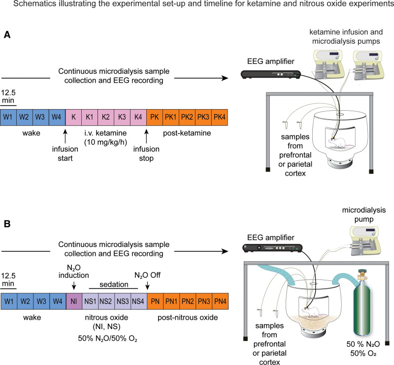
Figure 1.
Schematics illustrating the experimental setup and timeline for ketamine (A) and nitrous oxide (B) experiments. The EEG data and microdialysis samples from prefrontal and parietal cortices were collected simultaneously and continuously, but the microdialysis samples were collected in 12.5-min bins. Each colored box represents 1 microdialysis epoch. Samples were collected during freely moving baseline W condition, continuous K at 10 mg/kg/h, or PK recovery period. The data collection was performed similarly for the nitrous oxide cohort, with epochs corresponding to the W state, 50% NI, 50% NS, and PN recovery period. EEG indicates electroencephalogram; K, subanesthetic ketamine infusion; NI, nitrous induction; NS, nitrous sedation; PK, postketamine; PN, post-nitrous oxide; W, wake.

