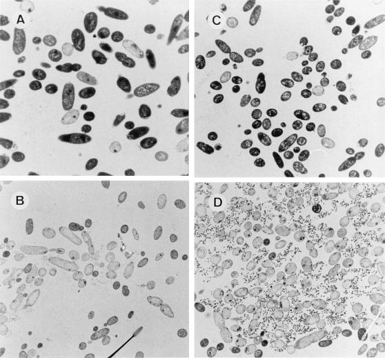
FIG. 5.
Transmission electron micrographs of ultrathin sections of P. putida CMC4 (A and B) and P. putida CMC12 (C and D) cells. A and C, noninduced cells growing in M9 minimal medium with glucose and 3-methylbenzoate. B and D, Induced cells 3 h after transfer to M9 minimal medium with glucose and IPTG. Magnification, ×4,000.

