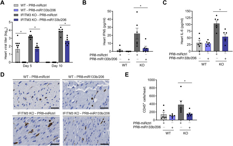
Fig. 4. PR8-miR133b/206 is attenuated in the heart in vivo.
WT and IFITM3 KO mice were intranasally infected with PR8-miR133b/206 or PR8-miRctrl (50 TCID50). (A to C) Mice were euthanized on day 5 or 10 after infection for TCID50 measurement of virus titers (D) or ELISA quantification of IFNβ (B) and IL-6 (C) and in the heart. Data points represent individual mice, and bars represent mean values. Error bars depict SD of the mean. Data points are from three independent experiments. Statistical comparisons were analyzed by ANOVA followed by Tukey’s post hoc test. *P < 0.05. (D) Mice were euthanized on day 10 after infection for histological analysis of CD45+ immune cell infiltration in the heart. Images shown depict areas of immune cell infiltration indicated by brown staining. Scale bars, 50 μm. (E) Whole-heart images were quantified for CD45+ cells using ImageJ. Data points represent individual mouse heart images, and bars represent mean values. Error bars depict SD of the mean. Comparisons were analyzed by ANOVA followed by Tukey’s post hoc test. *P < 0.05.

