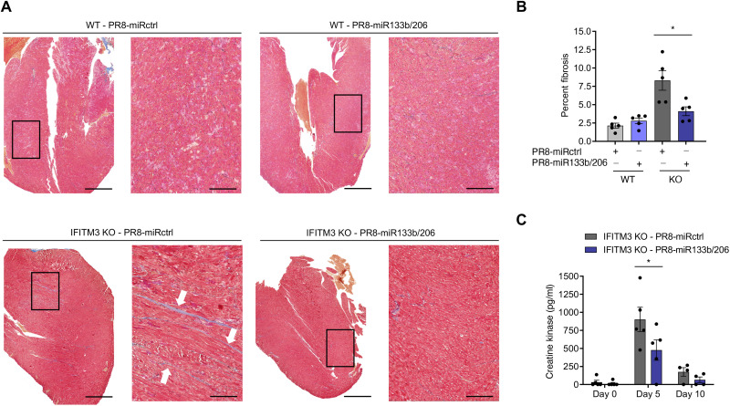
Fig. 5. Virus replication in the heart is necessary to induce fibrosis during infection.
WT and IFITM3 KO mice were intranasally infected with PR8-miR133b/206 or PR8-miRctrl (50 TCID50). (A) Hearts were collected on day 10 after infection, and sections were stained with Masson’s trichrome stain, in which blue staining is indicative of fibrotic collagen deposition. Histological processing and image acquisition were performed by the OSU Comparative Pathology and Mouse Phenotyping Core Facility on heart tissue samples provided by A.D.K. A representative heart section is shown for each genotype-virus combination. Boxed areas are regions magnified in the far-right images. Scale bars, 1 mm and 200 μm for the left and right images, respectively. (B) Percent fibrosis was calculated by quantifying ratio of blue pixel intensity to total pixel intensity for each heart section. Each point represents a heart from an individual mouse, and bars represent mean values. Error bars represent SD of the mean. Comparisons were analyzed by ANOVA followed by Tukey’s post hoc test. *P < 0.05. (C) Serum from IFITM3 KO mice was collected before infection and at days 5 and 10 after infection with PR8-miRctrl or PR8-miR133b/206 for ELISA quantification of creatine kinase. Data points represent individual mice, and bars represent mean values. Error bars depict SD of the mean. Comparisons were analyzed by ANOVA followed by Tukey’s post hoc test. *P < 0.05.

