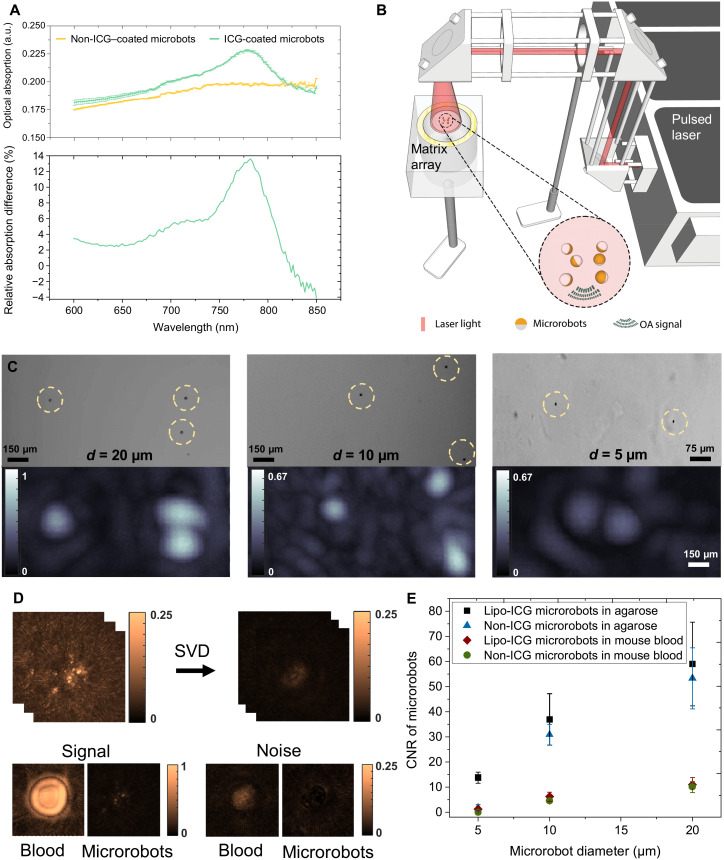
Fig. 2. Characterization of the magnetic microrobots with enhanced contrast for OAT.
(A) Optical absorbance spectra of the 10-μm-diameter microrobots coated with 50-nm layer of Au and 120-nm layer of Ni versus the microrobots additionally coated with Lipo-ICG. The latter generally exhibited higher optical absorption in the 600- to 820-nm wavelength range while further having a distinct peak at 780 nm, corresponding to the peak extinction of ICG. The bottom graph shows the excess of optical absorption contributed by Lipo-ICG coating in relation to pure Au/Ni coating. a.u., arbitray units. (B) Schematic drawing of the OAT imaging setup to perform single particle imaging. A phantom consisting of individual immobilized microrobots was uniformly illuminated from above with the generated optoacoustic signals captured by a spherical matrix array transducer. (C) Representative OAT and wide-field microscopy images of 20-, 10-, and 5-μm microrobots. The OAT image intensity is proportional to the size of the robots. However, because of the effective 150-μm resolution of the OAT imaging system, they appear much larger in the OAT reconstructions as compared to microscopic images. The scale bars refer to both the microscopy and OAT images. (D) For contrast-to-noise ratio (CNR) characterization with and without the presence of blood, a singular value decomposition (SVD) filter was applied to the raw data to render the background noise levels. It is shown that bloodless samples exhibit much lower background noise. (E) The estimated CNR in the 600- to 870-nm range for the two types of microrobots in the presence/absence of blood. Overall, the Lipo-ICG–coated microrobots exhibit slightly higher CNR than their counterparts only coated with Ni and Au. The CNR remains proportional to the microrobot size. Furthermore, the CNR is strongly diminished in the presence of blood.

