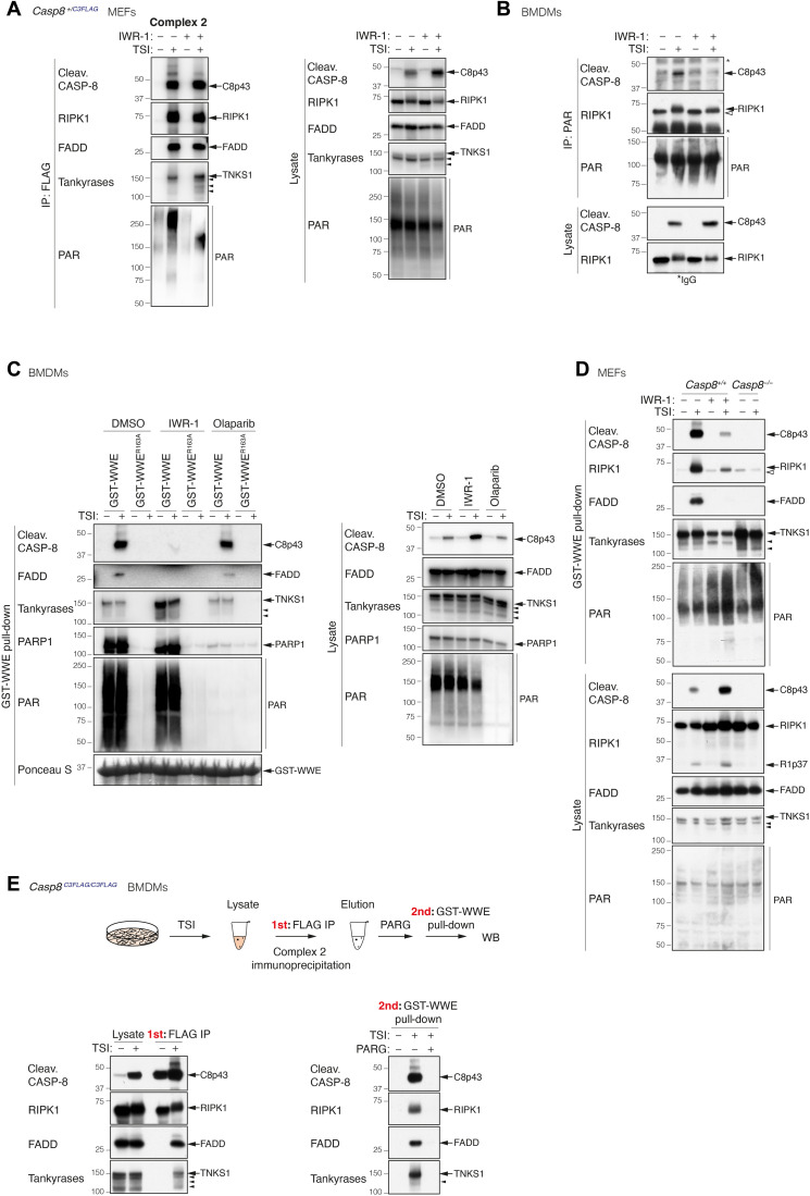
Fig. 2. Complex 2 is PARylated.
(A) Anti-FLAG immunoprecipitation of complex 2. Casp8+/C3FLAG MEFs were treated with TNF (100 ng/ml) + Smac mimetic (500 nM) + caspase inhibitor (5 μM; TSI) ± tankyrase inhibitor IWR-1 (10 μM) for 2 hours. (B) Anti-PAR (Trevigen 4335-MC-100) immunoprecipitation of complex 2. WT BMDMs were treated with TSI as in (A) ± tankyrase inhibitor IWR-1 (5 μM) for 1.5 hours. (C) GST-WWE pull-down of stimulated WT BMDMs lysates. Cells were treated with TNF (10 ng/ml) + Smac mimetic (250 nM) + caspase inhibitor (5 μM; TSI) ± tankyrase inhibitor IWR-1 (5 μM) or ± PARP1/2 inhibitor olaparib (1 μM) for 1.5 hours. Ponceau S staining of the purified proteins and their quantities is shown. (D) GST-WWE pull-down of stimulated MEFs lysates. Cells were treated as in (A). (E) Enrichment of PARylated complex 2 using GST-WWE in a sequential pull-down analysis. Casp8C3FLAG/C3FLAG BMDMs were treated with TSI [as in (A)], and complex 2 was immunoprecipitated using anti-FLAG M2 affinity beads. Immunoprecipitants were eluted with 3xFLAG peptides followed by ± PARG treatment at 37°C for 3 hours before GST-WWE pull-down. PAR chains were recognized by anti-PAR [poly/mono–ADP-ribose (E6F6A) rabbit monoclonal antibody no. 83732; CST] (A and C) or anti-PAR (MABC547; Sigma-Aldrich) (B and D). Filled arrowheads alone indicate potential tankyrase species. Double bands around 150 kDa in anti-tankyrase blots indicate full-length TNKS1 (upper band, 150 kDa) and an undefined TNKS1 isoform (lower band). Empty arrowheads alone denote unmodified RIPK1 that is purified nonspecifically by either sepharose anti-PAR (B) or sepharose GST-WWE. * indicate IgG chains.

