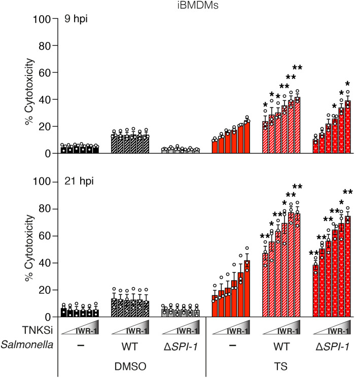
Fig. 7. Tankyrases protect against the cytotoxic effect of TNF under infection condition.
Level of cytotoxicity assessed by LDH assay. iBMDMs were infected with S. Typhymurium SL1344 WT or ΔSPI-1 (MOI: 2) and treated with TNF (10 ng/ml) + Smac mimetic (250 nM; TS) ± IWR-1 (250 and 500 nM and 1, 2, and 5 μM) at 3 hpi. Cytotoxicity was then assessed by LDH assay of cell supernatants collected at 9 and 21 hpi. Graph shows means ± SEM. n = 3 independent experiments. Comparisons were performed between TS-treated uninfected and S. Typhimurium SL1344 WT or ΔSPI-1-infected cells at each IWR-1 concentration with a Student’s t test whose values are denoted as *P ≤ 0.05 and **P ≤ 0.01.

