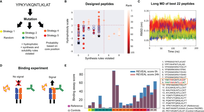
Figure 2.
Influenza peptide design for binding to a single allele. (A) Design strategies implemented: random mutations (strategy 1), mutations filtered by bioinformatic properties (strategies 2 and 3) and mutations filtered by MHC II binding motifs (strategies 4 and 5). (B) Hydrophobicity values, the number of synthesis rules violated and the average rank (scoring from 5 ns MD simulations) for the designed peptides. The thresholds (red dashed lines) used for selecting 22 peptides (blue circles) for long MD simulations. The original peptide is represented by a blue-filled circle. The controls are represented as black stars. (C) Cα RMSD to the starting structure of the 22 selected peptides during the long MD, colored by the mutation strategy panel (A). (D) Illustration of the Proimmune REVEAL binding assay. The peptide (yellow) binds the MHC II protein. When the components bind in the correct conformation, a positive signal is generated via a labelled antibody (green). (E) Experimental results of the REVEAL assay for the 17 designed peptides, the original sequence and two positive controls (PADRE and Influenza peptide PKYVKQNTLKLAT). The REVEAL binding score is a value between 0 and 100, which is determined by the comparison with a control T-cell epitope. The peptides are split into the five mutation strategies [color code as in panel (A)]. The scores are shown at 0 hours (blue bar) and after 24 hours (purple bar). The box highlights the two peptides with better binding than the reference sequence, with the modifications in bold, and colored in red and green to represent changes in the core and flanking regions, respectively.

