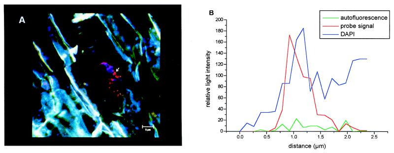
FIG. 3.
In situ detection of indigenous archaea on rice roots with rhodamine-labeled domain-specific oligonucleotide probe ARC915 (35). (A) The red dots in the center are coccoid archaeal cells that were 0.5 to 0.8 μm in diameter and specifically hybridized with probe ARC915. The photograph is an overlay resulting from three individual examinations, in situ hybridization with ARC915, DAPI staining, and autofluorescence of the plant tissue. The specificities of the hybridization signals were verified by measuring the relative signal intensities obtained from the oligonucleotide probe signal, the DAPI signal, and the autofluorescence signal, as shown for one optical cut in panel B (indicated by the arrow in panel A). The root cells are blue-green due to autofluorescence, and the dark areas correspond to the iron precipitates which often cover rice roots. The maximum distance between the archaeal cells and the root tissue was less than 12 μm, as determined by a z-series of optical sections, each 0.3 μm thick. Scale bar = 5 μm. (B) The three curves indicate the intensity of the oligonucleotide probe hybridization signal (red) in relation to the signal intensities of DAPI staining (blue) and autofluorescence of the plant tissue (green) for one optical cut with a high signal/noise ratio. The signal intensities were quantified by using the appropriate quantifying tools of the confocal laser scanning microscope.

