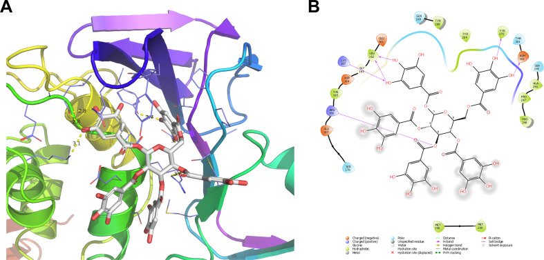
Fig. 2.
Docking model of PGG in SARS-CoV-2 PLpro. A Docking pose of PGG in the BL2 loop binding site of PLpro. B 2D ligand-protein interaction plot of PGG with SARS-CoV-2 PLpro. Docking was performed using the X-ray crystal structure of SARS-CoV-2 PLpro (PDB; 7JRN). The Glide score was −10.024 kcal/mol from the Schrödinger Glide extra-precision docking

