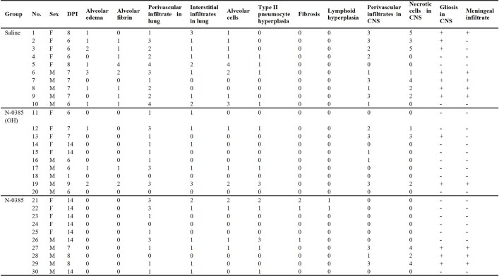
Extended Data Table 3.
Summary of histological findings
Associated with Fig. 4. For lung, scores were applied based on the percentage of each tissue type (alveolus, vessels, etc.) affected using the following criteria: (0) normal, (1) <10% affected, (2) 10–25% affected, (3) 26–50% affected, and (4) > 50% affected. For brain, histological scoring was assessed for perivascular inflammation using the most severely affected vessel and the following criteria: (0) no perivascular inflammation, (1) incomplete cuff one cell layer thick, (2) complete cuff one cell layer thick, (3) complete cuff two to three cells thick, and (4) complete cuff four or more cells thick. Necrotic cells in the neuroparenchyma were assessed per 0.237 mm2 field using the most severely affected area and the following criteria: (0) no necrotic cells (1) rare individual necrotic cells, (2) fewer than 10 necrotic cells, (3) 11 to 25 necrotic cells, (4) 26 to 50 necrotic cells, and (5) greater than 50 cells. DPI, days post-infection.

