FIGURE 4.
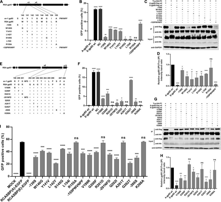
Identification of the key residues in the s3, s5, s8, and vr3 regions for the ALV-A binding receptor and invading cells. (A,E) Schematic representation of amino acid substitutions in s3 and s5 or s8 and s12. (B,F) DF-1 cells were incubated with mutant gp85 proteins with amino acid substitutions in s3 and s5 or s8 and s12 and subsequently infected with RCASBP (A)-EGFP. The percentage of GFP-positive cells was measured by FACS for blocking analysis. (C,G) The interaction of the mutant gp85 protein with amino acid substitutions in s3 and s5 or s8 and s12 with Tva-HA-Fc. (D,H) The gray scale value level of gp85 (IP)/Tva (IP) was calculated. (I) DF-1 cells transfected with recombinant RCASBP (A/E)-EGFP vectors with mutations V138N, −151N, Δ214–215, R222G, or R223G were detected for the percentage of GFP-positive cells by FACS 5 days posttransfection. *P < 0.05, **P < 0.01, ***P < 0.001, ****P < 0.0001.

