
Figure 4.
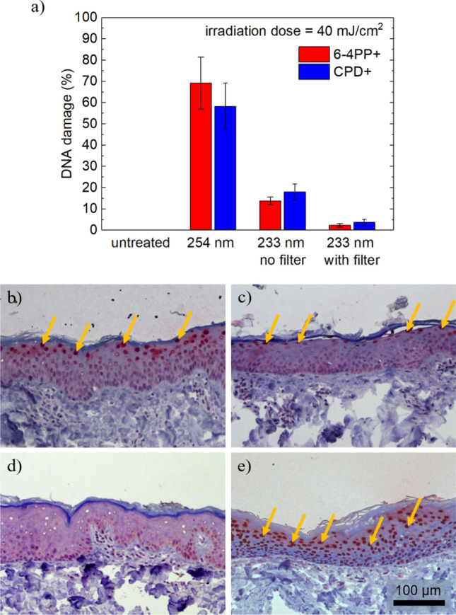
(a) DNA damage for irradiated porcine skin using 254 nm and 233 nm light sources with 40 mJ/cm2. Untreated skin served as control. The mean values of the epidermal DNA damage in % are calculated from at least three experiments and the error bars show the standard error of the mean. (b)–(e) Histologic images HE and CPD stained porcine skin after irradiation using the far-UVC LED irradiation system without (c) and with filter (d), in comparison, untreated skin (e) and skin after irradiation with near-UVC radiation at 254 nm (f). Arrows mark CPD positive cells. The scale bar is 100 μm.