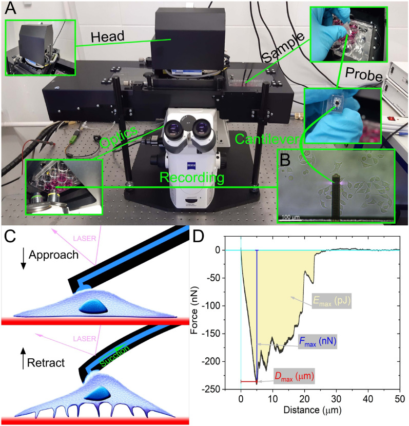
Figure 1.
Schematic representation of the measurement setup and procedure. On the FluidFM (A), living cell cultures can be observed with an optical microscope (see insert B where the cantilever is clearly visible). The large area sample stage under the measurement head allows multiple cell targeting in cm scale areas. During SCFS recording, cells are approached with the hollow FluidFM cantilever, which pauses upon contact with the targeted cell, and suction (vacuum) is applied to attach the cell to the aperture, after which the cantilever is retracted from the substrate (C). The SCFS measurement process yields the characteristic FD curves, presenting the primary parameters Fmax, Emax, and Dmax (D).

