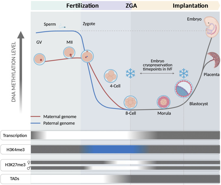
FIGURE 1.
Representation of the human embryo epigenomic state at 8-cell and at blastocyst stage, both common timepoints for application of cryopreservation within clinical ART. Fertilization releases embryo reprogramming by inducing a wide remodeling along different epigenetic layers. The 8-cell stage and the blastocyst stage are illustrated to represent their very different epigenomic states of their embryonic cells. A sharp demethylation takes place in both, the sperm and the oocyte genomes, with this last showing a more paused progression. The demethylation becomes maximum at 8-cell stage, coinciding with the ZGA. BLUE indicates an unusual positioning of at H3K4me3 on CpG rich regions at promoters of transcribed genes and also positioned at distal CpG rich and hypomethylated regions until 8-cell stage. Intriguingly, H3K4me3 lacks at CpG rich promoter regions of developmental and differentiation related genes (Xia et al., 2019). H3K27me3 is progressively erased after fertilization and completely absent during ZGA, with a slight delay in the maternal genome. An asymmetric distribution of H3K27me3 has been shown in blastocyst by its positioning in ICM cells at distal cis-regulatory regions of developmental genes (Xia et al., 2019). TADs formation arise from 8-cell and according to previous observations is dependent on ZGA onset, which surrounds 8-cell stage (Chen et al., 2019). GREY areas show a common pattern observed in differentiated cells; WHITE indicate a loss of activity; BLUE indicates a different role than that of differentiated cells.

