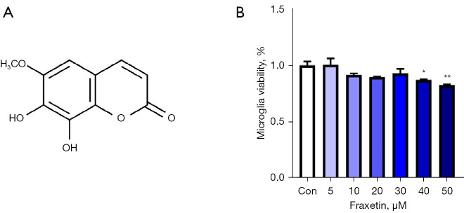
Figure 1.
Effects of fraxetin on the viability of primary microglia. (A) Chemical structure of fraxetin. (B) Primary microglia were treated with different concentrations of fraxetin (5, 10, 20, 30, 40, or 50 µM). After 24 hours, a CCK-8 assay was performed to detect the viability of the microglia. Control group n=6, other groups n=3. The values represent the mean ± SEM. *P<0.05, **P<0.01 compared with the control group. CCK-8, cell counting kit-8; SEM, standard error of the mean.

