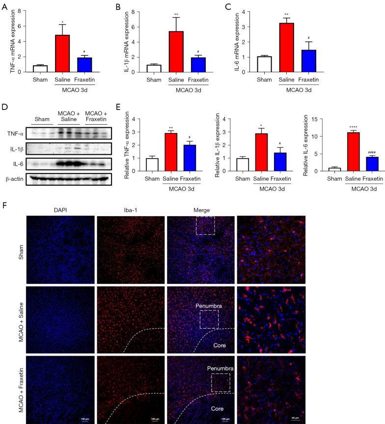
Figure 5.
Fraxetin attenuated proinflammatory cytokine expression and microglial activation after MCAO. (A-C) TNF-α (A), IL-1β (B), and IL-6 (C) mRNAs of the penumbra tissues were quantified using RT-PCR. n=3–6 per group. (D-E) TNF-α, IL-6, and IL-1β proteins in the penumbra tissues were evaluated using western blotting (D) and quantified (E). n=3 per group. (F) Representative images of tissue sections from the ischemic penumbra were collected 3 days after MCAO, and stained with Iba1 and DAPI. The inset on the right side shows a digitally magnified field of cells from the image. Scale bars: 100 and 40 µm. The values represent mean ± SEM. *P<0.05, **P<0.01 and ****P<0.0001 vs. sham group. #P<0.05, ####P<0.0001 vs. MCAO-saline groups. MCAO, middle cerebral artery occlusion; TNF, tumor necrosis factor; IL-1β, interleukin-1 beta; IL-6, interleukin-6; RT-PCR, real-time polymerase chain reaction; DAPI, 4',6-diamidino-2-phenylindole; SEM, standard error of the mean.

