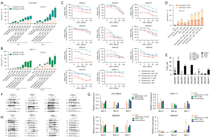
Figure 2.
Apoptosis rates and gene and protein expression of apoptosis-related molecules in AML cells treated with venetoclax and HHT. (A,B) The apoptosis rates of AML cell lines treated with venetoclax and HHT alone or in combination for 8 h and 24 h [(A) OCI-AML3; (B) MV4-11]. (C) The viable cell ratio of primary AML samples after treatment with different concentrations of venetoclax, HHT, or their combination. (D) The IC50 value of venetoclax based on the viable cell ratio and the second-generation sequencing results of patient bone marrow samples. (E) The apoptosis rate of AML#01 primary cells treated with venetoclax and HHT alone or in combination for 24 h. (F) Changes in apoptosis-related proteins detected by WB. (G) Changes in Bcl-2 family apoptotic regulatory molecules detected by qRT-PCR. (H) Changes in Bcl-2 family apoptotic regulatory proteins detected by WB. *, P<0.05; **, P<0.01; ***, P<0.001, compared with the control group. HHT, homoharringtonine; AML, acute myeloid leukemia; WB, Western blotting; qRT-PCR, quantitative reverse transcription-polymerase chain reaction; CI, combination indices.

