. 2022 Apr 26;305(1):46–53. doi: 10.1148/radiol.220543

Figure 3:

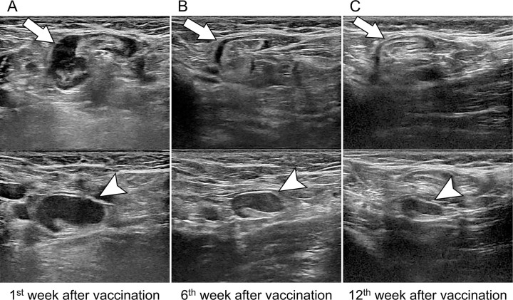
Images in a 43-year-old asymptomatic woman without breast cancer and unilateral left axillary adenopathy after messenger RNA COVID-19 vaccination. Axial US images of the left axilla show two enlarged lymph nodes (arrow, top; arrowhead, bottom). (A) US scan obtained within the 1st week of BNT162b2 vaccination shows the cortex measuring up to 7.7 mm (arrowhead). (B, C) Follow-up US images obtained within 6 weeks (B) and 12 weeks (C) show decreased axillary lymphadenopathy of variable degrees, with the cortex measuring up to 5.7 and 3.4 mm (arrowhead), respectively.

Images in a 43-year-old asymptomatic woman without breast cancer and unilateral left axillary adenopathy after messenger RNA COVID-19 vaccination. Axial US images of the left axilla show two enlarged lymph nodes (arrow, top; arrowhead, bottom). (A) US scan obtained within the 1st week of BNT162b2 vaccination shows the cortex measuring up to 7.7 mm (arrowhead). (B, C) Follow-up US images obtained within 6 weeks (B) and 12 weeks (C) show decreased axillary lymphadenopathy of variable degrees, with the cortex measuring up to 5.7 and 3.4 mm (arrowhead), respectively.