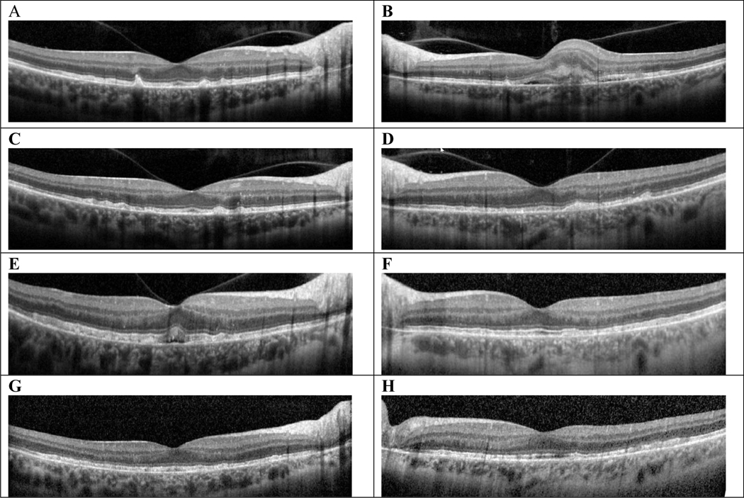
Figure 2.
OCT stack with a horizontal cut through the foveal center at each time point showing evolution of both eyes. A and B, At initial visit, this 47-year-old woman had drusen and SDDs OU, and subretinal fluid with subretinal hyperreflective material OS consistent with CNV OS. C and D, 2 months after initial presentation, scattered RPE elevations and SDDs OU with regressed CNV OS. E and F, 2 years after initial presentation, there was new subretinal fluid OD, RPE/EZ irregularities, diffuse RPE elevations, EZ band irregularities OU. G and H, 8 years after presentation, diffuse RPE/EZ band irregularities. CNV indicates choroidal neovascularization; EZ indicates ellipsoid zone; OCT, optical coherence tomography; OD, right eye; OS, left eye; OU, both eyes; RPE, retinal pigment epithelium; SDD, subretinal drusenoid deposits.

