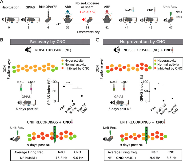
Fig. 9.
Recovery from tinnitus-behavior when decreasing activity of CaMKII α+ DCN neurons acutely but not if activity was chemogenetically reduced during the noise exposure. A Schematic outline highlighting the hypothetical difference between experiments of recovery in tinnitus-like animals (B) and animals where the aim was to prevent tinnitus (CNO administered before the noise exposure, second set of experiments as shown in C, red font). B Top, schematic drawing of the DCN fusiform layer showing hypothetical hyperactive neurons following noise exposure. Middle, CNO improved gap detection. Bottom, CNO administration causes a reduction in average firing frequency that may be due to inhibiting hyperactive DCN neurons. C Top, schematic drawing showing the DCN fusiform layer chemogenetically inhibited during the noise exposure; however, some neurons are probably not affected by CNO and may still render some neurons hyperactive. Middle, acute CNO exposure does not improve gap detection in mice administered CNO during the noise exposure. Bottom, unit recordings in animals administered CNO during the noise exposure shows that acute additional CNO administration reduces the overall firing frequency less dramatically compared to “B,” suggesting units expressing hM4Di receptors were not hyperactivated during the noise trauma

