FIGURE 6.
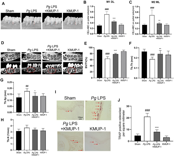
KMUP-1 protects against periodontal bone loss induced by PgLPS. The periodontitis was induced by repeated injection of PgLPS into gingiva. (A) Representative micro-CT images of palatal maxilla of experimental groups. The distance between cementoenamel junction (CEJ) to alveolar bone crest (ABC) at (B) first maxillary molar (M1, as denoted by blue lines) of distolingual (DL) and (C) second maxillary molar (M2, as denoted by red lines) of mesiolingual (ML) was measured 3 weeks after KMUP-1 treatment. (D) Representative longitudinal- and cross-sectional micro-CT images of the first and second molar in the maxilla. Circles indicate region of interest. The volumetric parameters, including (E) bone volume/tissue volume (BV/TV), (F) trabecular thickness (Tb.Th), (G) trabecular separation (Tb.Sp), and (H) trabecular number (Th.N) were measured. (I) Representative image of TRAP stain in maxillary alveolar bone sections. Arrows indicate TRAP+ cells. (J) Statistical analysis of osteoclast (TRAP+ cells) density in the maxillary alveolar bone. Scale bars: 100 μm n = 6. ##p < 0.01, ###p < 0.001 compared with sham. **p < 0.01, ***p < 0.001 compared with PgLPS.

