Fig. 2.
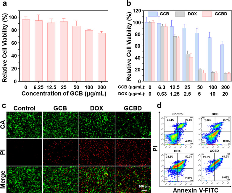
The analysis of cell death pathway. a Relative cell viability of HUVEC cells after incubation with GCB NDs at different concentrations (0–200 µg/mL) for 24 h. b Relative cell viability of 4T1 cells after incubation with GCB NDs (0–200 µg/mL), DOX (0–20 µg/mL) or GCBD NPs (0–20 µg/mL of DOX) for 24 h. c Confocal imaging of calcein-AM/PI stained 4T1 cells after incubation with different solutions for 24 h. d The flow cytometry analysis of 4T1 cells after incubation with different solutions for 24 h, showing more significant apoptosis in GCBD NPs-treated cells than other groups (2.5 µg/mL for DOX)

