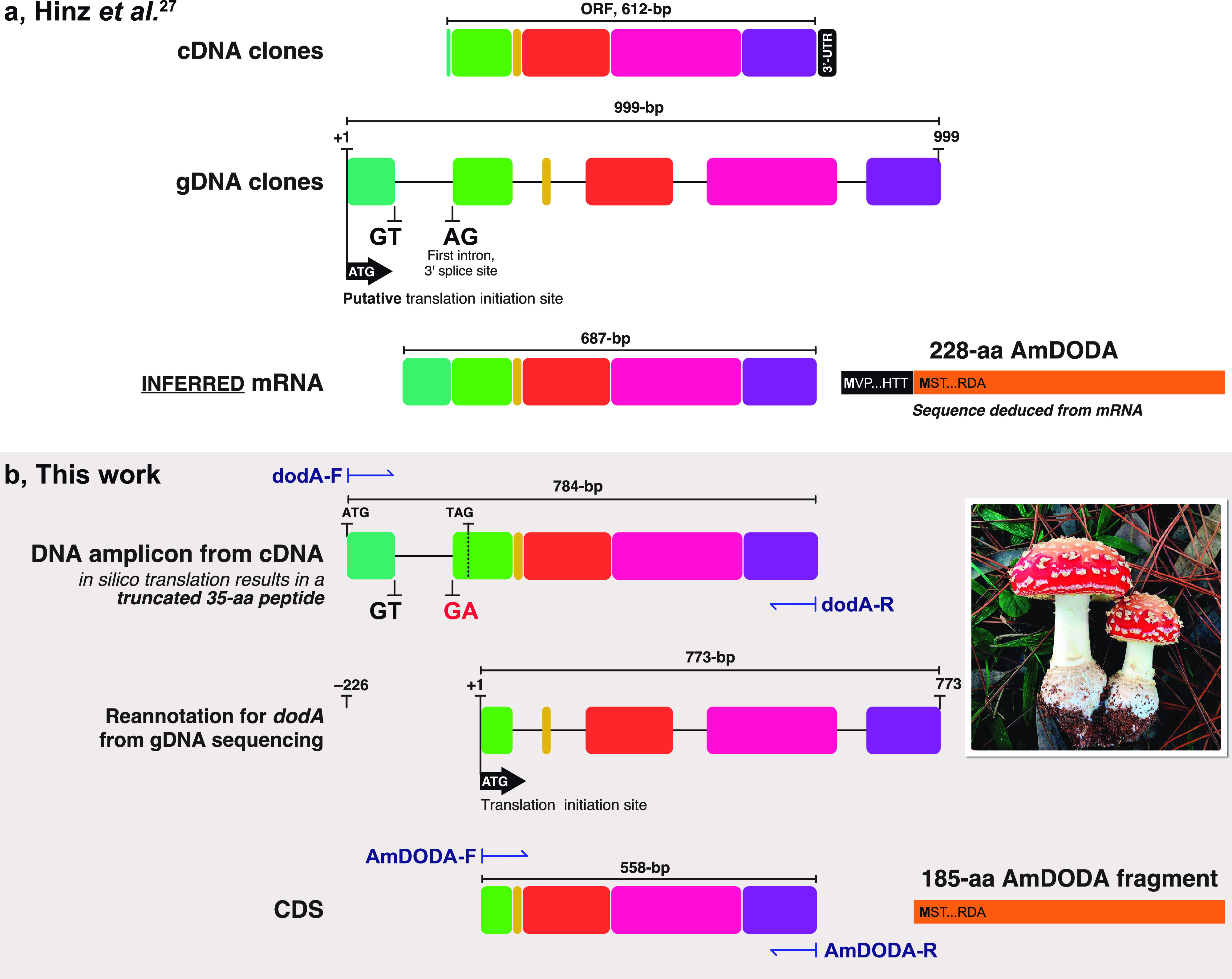
Figure 2.

Comparison of the study reported by Hinz and coauthors27 and this work. (a) Structures of cDNA and gDNA clones isolated by Hinz and coauthors and the inferred 687-bp mRNA. Introns are presented as lines and exons as colored boxes; this schematic representation was created from the sequences reported in ref (27). The 228-aa AmDODA is represented by the black and orange bar showing the initial and final amino acid triads. The sequence was deduced by Hinz and coauthors from the mRNA and is presented in Figure S3. (b) DNA amplicon obtained by PCR amplification of the CDS for AmDODA from cDNA samples of Eurasian A. muscaria specimens collected in São Paulo, Brazil (inset photo; credit: Prof. C. V. Stevani). The “GA” replacing the canonical “AG” reported by Hinz and coathors leads to the retention of the first intron. A TAG stop codon indicates the 3′-end of the ORF used for the in silico translation of the 784-bp DNA fragment, which would result in a 35-aa truncated polypeptide. The structure of the dodA gene, as determined from DNA sequencing of gDNA amplicons, is shown and the transcription initiation site is indicated. The 558-bp CDS encoding the 185-aa AmDODA described in this work is presented. PCR primer sites are indicated using dark blue harpoon arrows. The complete sequence of the dodA gene is presented in Figure S2.
