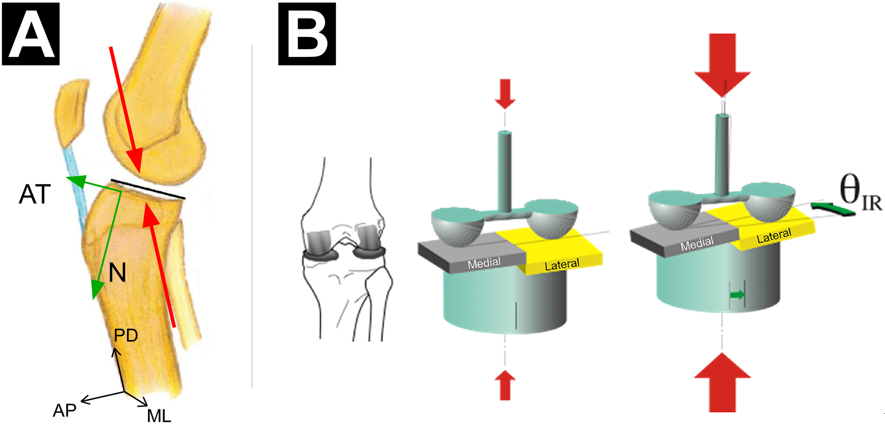
Figure 4.

(A) Sagittal-plane view of a right knee illustrating how an axial knee compression force can induce coupled anterior tibial translation because of the geometry of the articulating surfaces of the tibiofemoral joint. As a result of the posterior-inferior-directed slope of the lateral and medial tibial plateau, the knee compression force (red arrows, acting along the proximal-distal (PD) axis of the tibia) has a component that induces an anterior tibial shear force (green arrow, AT), in addition to the normal force (green arrow, N, force component perpendicular to the surface of the tibial plateau). (B) Postero-superior three-quarter view of a right knee illustrating how a large axial knee compression force (large red arrows) can induce coupled internal tibial rotation. As a result of the steeper posterior-inferior-directed slope of the lateral tibial plateau in comparison with that of the medial tibial plateau, the lateral femoral condyle essentially pushes the lateral tibial plateau anteriorly more than the medial femoral condyle pushes the medial tibial plateau thereby causing internal tibial rotation (IR). Reproduced from Wojtys et al.155
