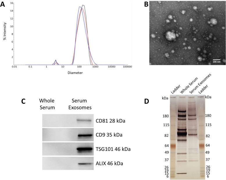
Fig. 4.
Characterization of serum exosomes. A Dynamic light scattering (DLS) results performed in triplicate indicating an average diameter of 164 nm. B Transmission electron microscopy (TEM) demonstrating a heterogenous size mixture of discrete vesicles ranging from about 30 – 200 nm. C SDS-PAGE analysis of whole serum and serum exosome protein patterns visualized by silver staining. D Western blot results showing intense CD9, CD81, TSG101, and ALIX (exosome protein markers) signals in the serum exosomal fraction, but not in the whole serum

