. 2019 Oct 11;9(56):32499–32509. doi: 10.1039/c9ra06614d

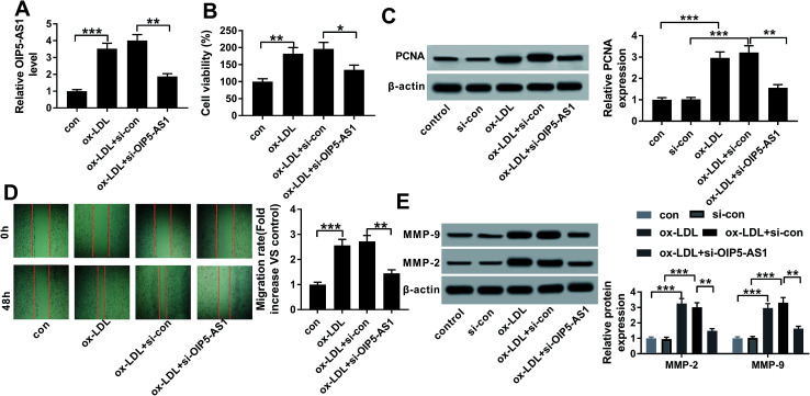
Fig. 2. Role of OIP5-AS1 in cell proliferation and migration in hVSMCS. hVSMCs were transfected with siRNA against OIP5-AS1 (si-OIP5-AS1) or its control (si-con), followed by 100 μg mL−1 of ox-LDL treatment or not. (A) RT-qPCR detected OIP5-AS1 levels at 24 h. (B) Cell counting kit-8 (CCK-8) measured cell proliferative capacity at 0 h, 24 h, 48 h and 72 h. (C) Western blotting examined expression of proliferating cell nuclear antigen (PCNA) at 24 h. Protein bands of PCNA was quantified by densitometry and normalized to the level of β-actin. (D) Wound healing assay determined the ability of cell migration at 48 h. Migration rate was fold change of migration distance versus original distance. (E) Western blotting examined expression of matrix metalloproteinase (MMP)-2 and MMP-9 at 24 h. Protein bands of MMP-2/9 was quantified by densitometry and normalized to the level of β-actin. Data represent mean ± SEM. **P < 0.01 and ***P < 0.001 according to one-way analysis of variance (ANOVA).

Fig. 2