
FIGURE 1.
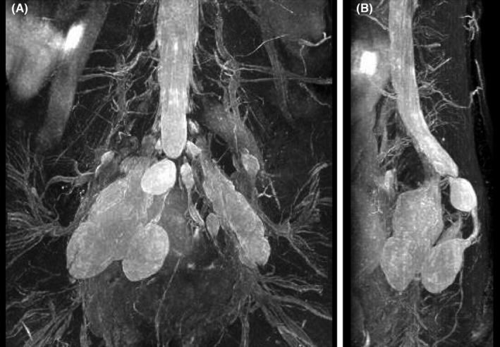
Magnetic resonance imaging findings (magnetic resonance myelography) at 23 weeks of gestation. Spindle‐shaped, bilateral cysts protruding into the pelvic cavity are shown via (A) coronal and (B) lateral views
Official websites use .gov
A
.gov website belongs to an official
government organization in the United States.
Secure .gov websites use HTTPS
A lock (
) or https:// means you've safely
connected to the .gov website. Share sensitive
information only on official, secure websites.
Magnetic resonance imaging findings (magnetic resonance myelography) at 23 weeks of gestation. Spindle‐shaped, bilateral cysts protruding into the pelvic cavity are shown via (A) coronal and (B) lateral views