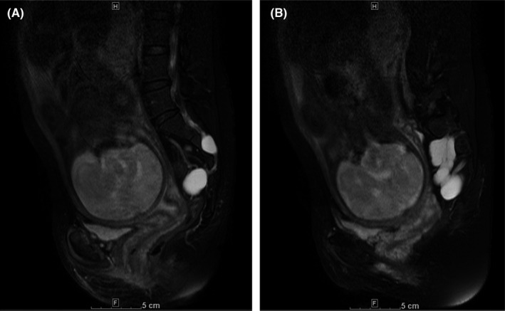
FIGURE 2.

Magnetic resonance imaging findings (sagittal fat‐suppressed T2‐weighted imaging) at 34 weeks of gestation. (A) The minimum distance between the cyst wall and posterior surface of the pubis was 100 mm and (B) Tarlov cysts adjacent to the baby's head on the sagittal‐section 2 cm to the right of the midline
