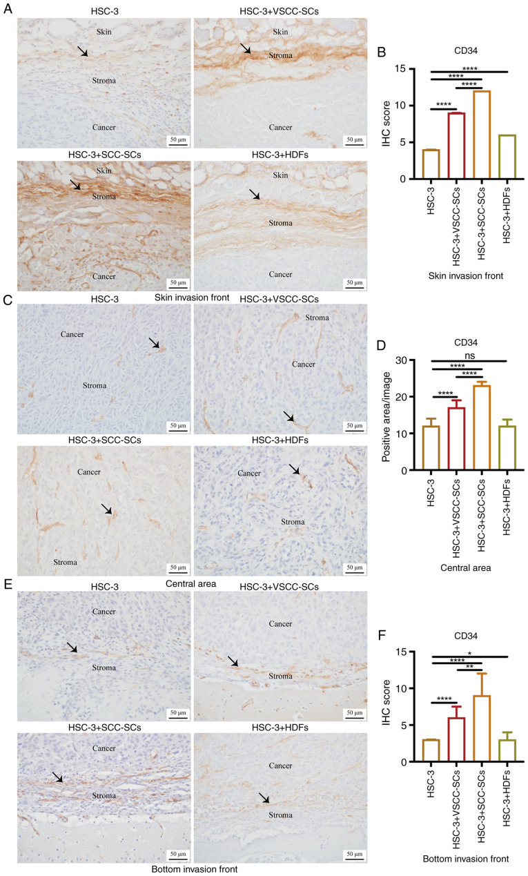
Figure 4.
Effects of VSCC-SCs, SCC-SCs and HDFs on the MVD in the tumor microenvironment of OSCC following crosstalk with HSC-3 cells in vivo. (A, C and E) Immunohistochemical staining was used to examine the expression of CD34 in the (A) skin invasion front, (C) central area and (E) bottom invasion front. Black arrows indicate vessel structure. (B, D and F) Quantification of CD34 expression in the (B) skin invasion front, (D) central area and (F) bottom invasion front in the different groups. Data are presented as the median and IQR, n=4. Statistical analysis was performed using the Kruskal-Wallis test followed by Dunn's test.*P<0.05, **P<0.01 and ****P<0.0001. ns, not significant (P>0.05); VSCC-SCs, verrucous squamous cell carcinoma-associated stromal cells; SCC-SCs, squamous cell carcinoma-associated stromal cells; HDFs, human dermal fibroblasts.

