
Figure 1. Prostate cancer educates MLO-Y4 osteocytes to promote C4-2B prostate cancer cell proliferation, migration and invasion.
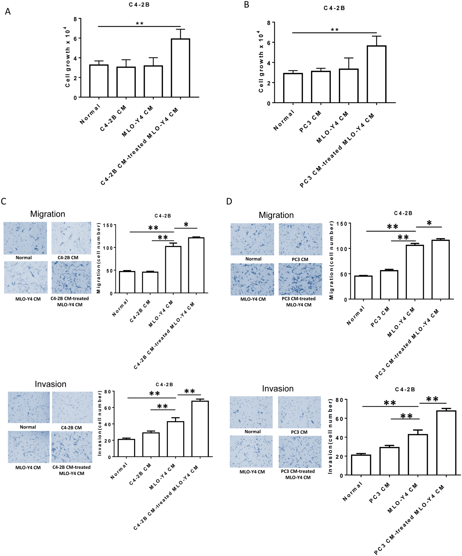
Conditioned-media (CM) from both C4-2B and PC3 prostate cancer cells was made as described in methods. CM was added as described to a final concentration of 50%. (A). Plain media, C4-2B CM, MLO-Y4 CM and C4-2B CM-treated MLO-Y4 cell CM were added to C4-2B cells (1.5×10⁴ per well). After 48 hours, cell numbers were counted using a hemocytometer. (B). Plain media, PC3 CM, MLO-Y4 CM and PC3 CM-treated MLO-Y4 cell CM were added to C4-2B cells (1.5×10⁴ per well). After 48 hours, cell numbers were counted using a hemocytometer. (C). Migration (upper figures) and invasion (lower figures) were assessed using a transwell assay. C4-2B cells (1.5×10⁵ per cells) were treated with plain media, C4-2B CM, MLO-Y4 CM and C4-2B CM-treated MLO-Y4 cell CM for 24 hours. The membrane was stained using differential Quick staining kit and photographed under light microscopy (20x). The numbers of migrating (no Matrigel on membrane) and invading (Matrigel present on membrane) cells were counted in five random fields for each insert. (D). Migration (upper figures) and invasion (lower figures) were assessed using a transwell assay. C4-2B cells (1.5×10⁵ per cells) were treated with plain media, PC3 CM, MLO-Y4 CM and PC3 CM-treated MLO-Y4 cell CM for 24 hours. The membrane was stained using differential Quick staining kit and photographed under light microscopy (20x). The numbers of migrating (no Matrigel on membrane) and invading (Matrigel present on membrane) cells were counted in five random fields for each insert. Data are shown as the mean±SD of 3 independent experiments. * P < 0.05; ** P < 0.01.