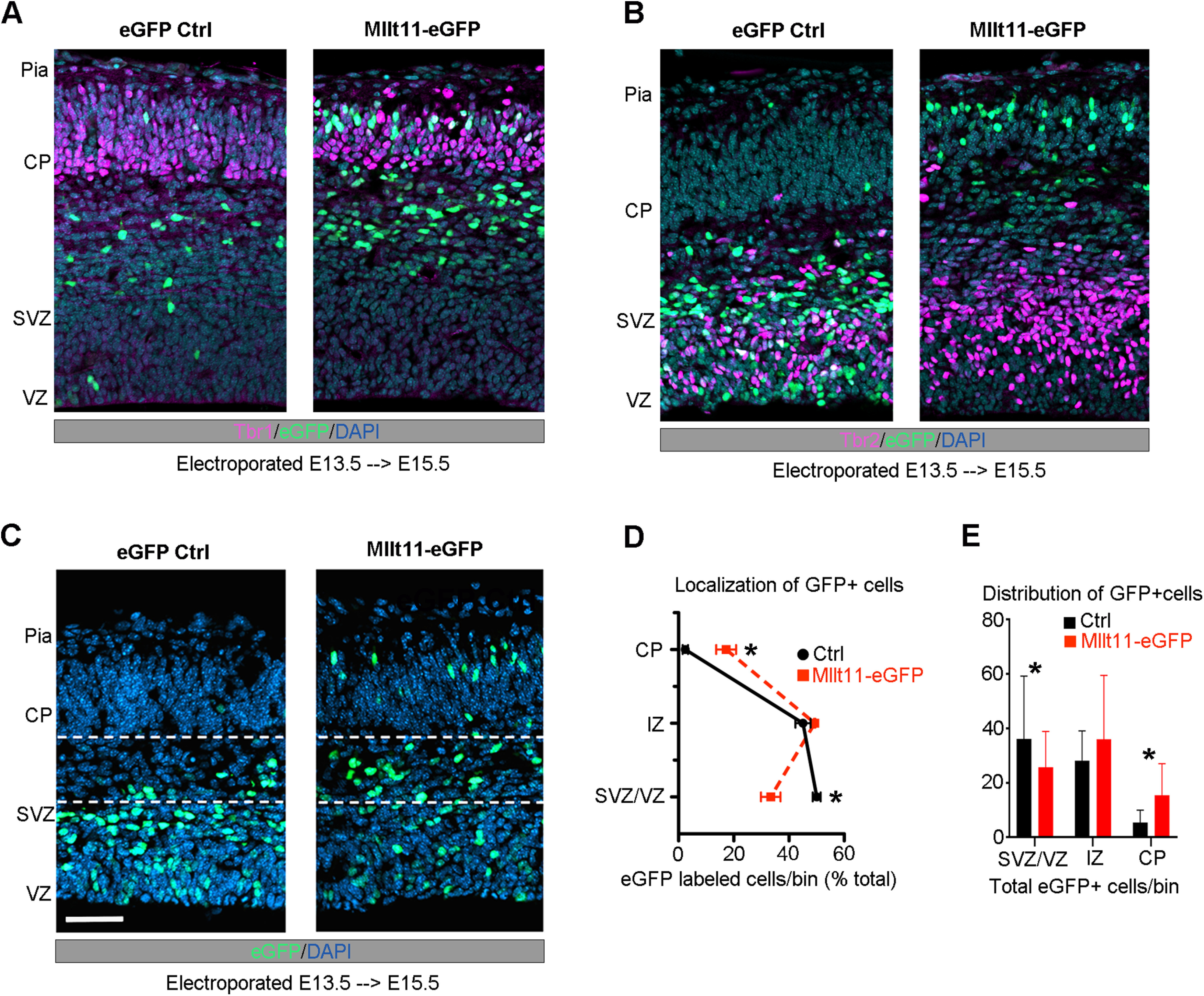
Figure 4.

Mllt11 overexpression promoted migration into the CP. A, B, E15.5 coronal cortical sections after electroporation at E13.5 wither either a control eGFP (left) or Mllt11-ires-eGFP (Mllt11-eGFP) bicistronic plasmid (right). B, Mllt11-eGFP electroporation promoted migration into the CP, identified by Tbr1 staining. C, Control GFP (eGFP-control) electroporated cells remained mostly within the SVZ/VZ, identified by Tbr2 staining. D, E, Localization of eGFP-control and Mllt11-eGFP+ cells quantified as a percentage of total eGFP+ cells (D), and as the distribution of total eGFP+ cells per fetal cortical layer bin (E). Student's t test with Welch's correction; N = 3 Mllt11-eGFP, N = 4 eGFP controls. Data presented as mean ± SD; *p ≤ 0.05. Scale bar: 25 μm (A). CP, cortical plate; IZ intermediate zone; SVZ, subventricular zone; VZ, ventricular zone.
