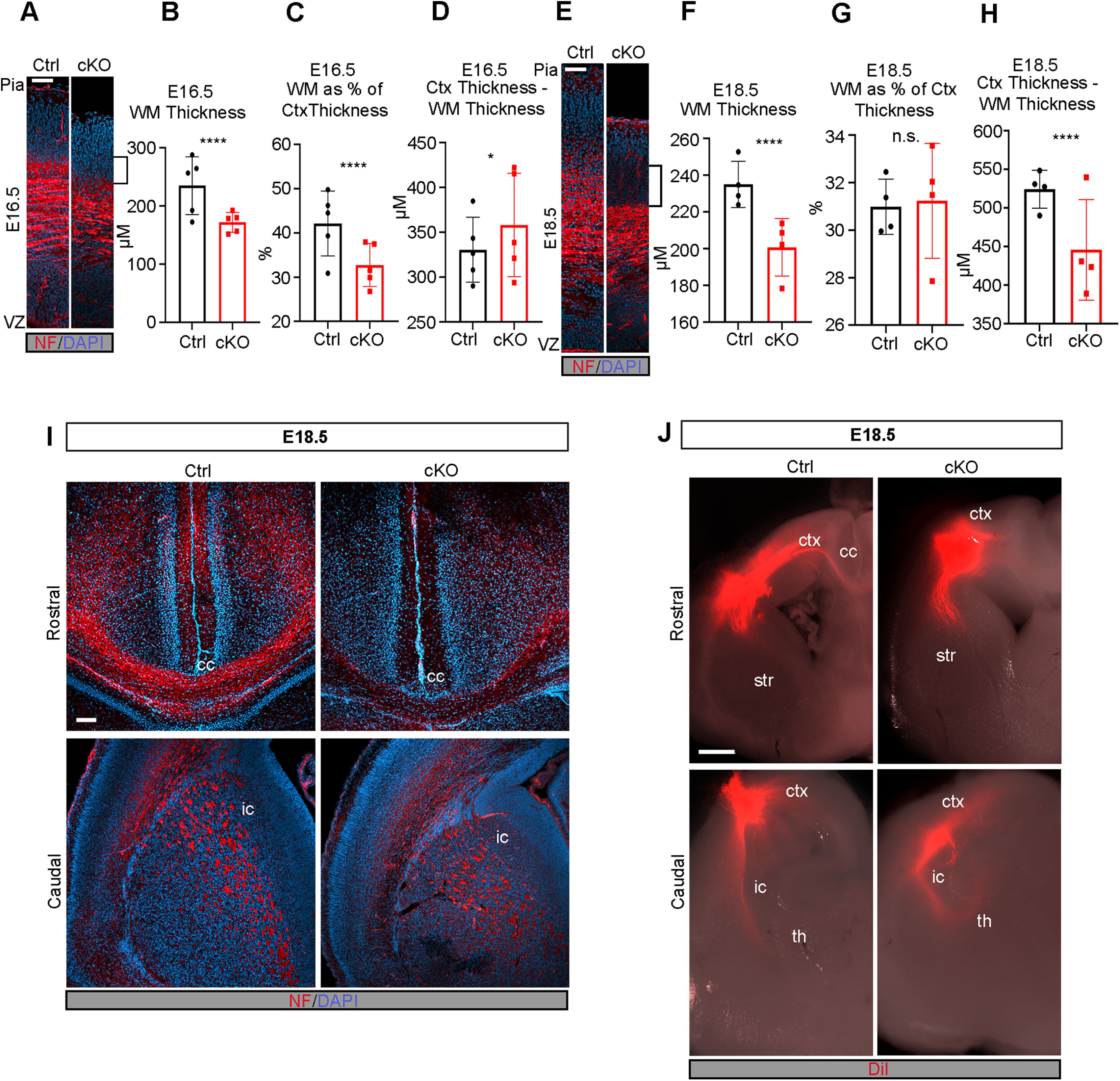
Figure 5.

Formation of WM tracts and callosal projections is impaired in the Mllt11 cKO cortex. A, Image of cortical WM tracts labeled with neurofilament at E16.5. Brackets indicate decreased WM. B, C, Quantification of cortical WM thickness (B) and WM thickness as a proportion of total cortical thickness (C) showed significant decrease in WM in cKOs relative to controls at E16.5. D, Quantification of cortical thickness subtracted from WM tract thickness showed a slight increase in Mllt11 cKO mutants compared with controls at E16.5. E, Image of cortical WM tracts labeled with neurofilament at E18.5. Brackets show decreased WM staining area. F–H, Quantification of cortical WM thickness (F), WM thickness as a proportion of total cortical thickness (G), and cortical thickness subtracted from WM tract thickness (H) all showed significant decreases in Mllt11 mutants compared with controls at E18.5. I, J, Coronal sections of E18.5 cortices at rostral (upper panels) and caudal (lower panels) axial levels labeled with neurofilament (I) or traced with DiI (J). I, Neurofilament labeling of the corpus callosum was significantly decreased in cKO compared with controls but labeling of the internal capsule was unaffected. J, DiI labeling was absent in the corpus callosum of cKO slices while control cortices displayed crossing fibers labeled by DiI. Rostrally, the internal capsule was traced comparably in control and cKO cortices. Student's t test with Welch's correction, (A–D) N = 5, (E–H) N = 4, (J) N = 3. Data presented as mean ± SD; ****p ≤ 0.0001. Scale bar: 200 μm (A) and 50 μm (B). ctx, cortex; cc, corpus callosum; str, striatum; th, thalamus; ic, internal capsule; VZ, ventricular zone; WM, white matter.
