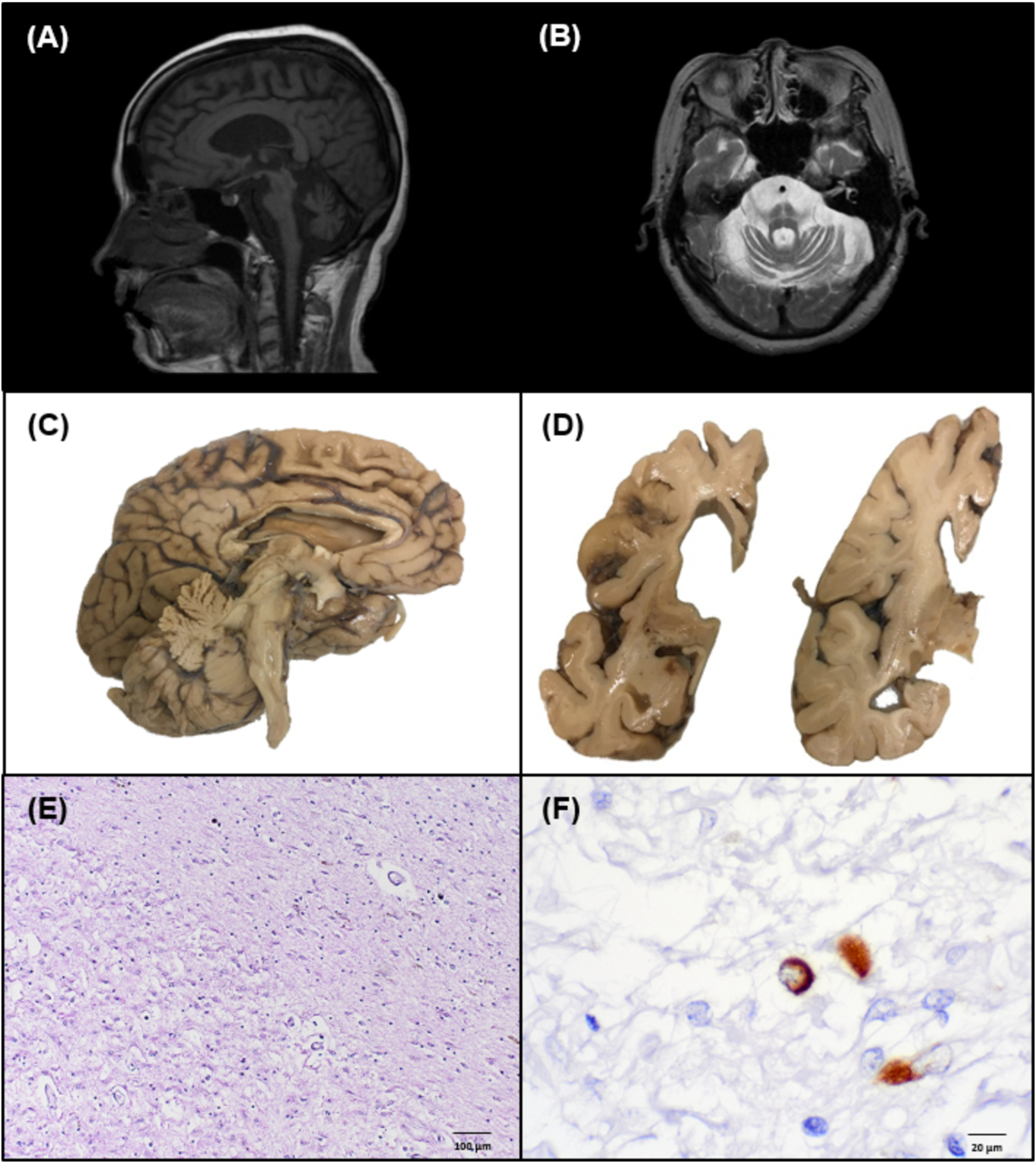
Figure 1. Radiologic, gross anatomic, and histologic neurodegenerative features of a brain with longstanding atypical MSA.

(A) Midsagittal T1-weighted MRI showing olivopontocerebellar atrophy. (B) Axial T1-weighted MRI showing cerebellar and pontine atrophy with increased T2 signal characteristic of the “hot cross bun sign” within the pons. (C) Mid-sagittal view of left hemibrain exhibiting olivopontocerebellar atrophy, medial frontal lobe atrophy, and cingulate gyrus atrophy. (D) Coronal view of left hemibrain highlighting basal ganglia, hippocampal atrophy, and hydrocephalus ex vacuo. (E) H&E stained section of the striatum demonstrating severe neuron loss, rarefaction, and gliosis. (F) Rare glial cytoplasmic inclusion identified on α-synuclein staining (81A IHC).
